Neuroinformatics Core (NIC)
Neuroreceptor Autoradiography Lab (HAL)
Short-lived-radioisotope Autoradiography and Radiometabolites Lab (SAL)
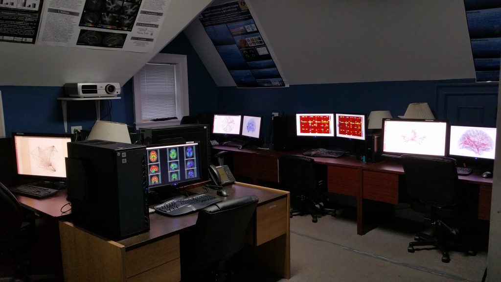
TNL-NIC has a dedicated 240-core computer cluster to process complex algorithms for analysis of post-mortem data as well as in vivo PET, MRI and genetics data. The computational environment has a dedicated imaging interface to facilitate the access to non-imaging experts, such as physicians, pharmacists or biologists. NIC computer cluster is the heart of all activities at TNL. Due to its great processing capabilities, NIC-TNL cluster permits the use of complex and innovative imaging techniques to validate novel in-vivo techniques using gold standard post-mortem data. In addition, TNL is connected to the supercomputer Consortium Laval UQAM McGill and Eastern Quebec-based in McGill University (CLUMEQ).
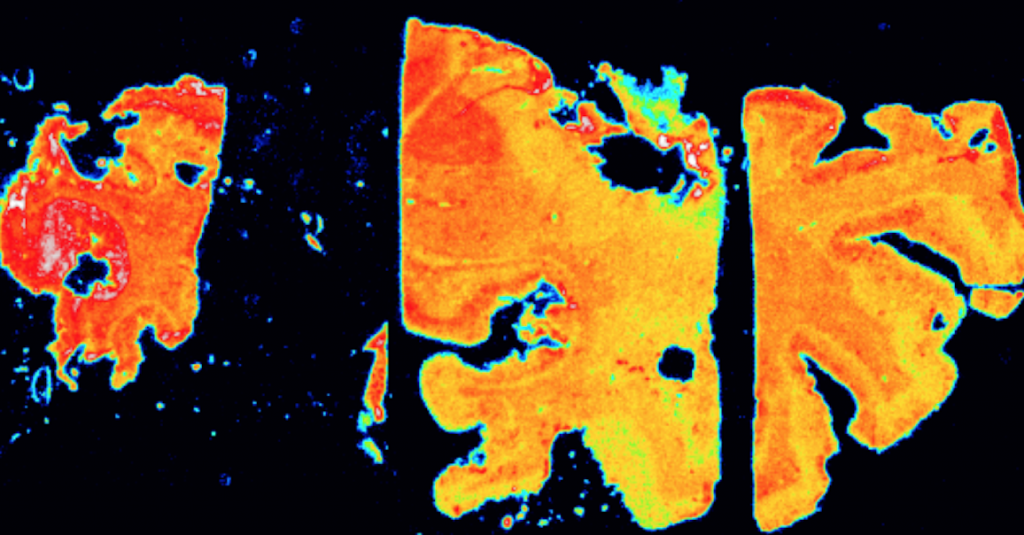
HAL is a CFI-funded post-mortem imaging laboratory for autoradiography capable of imaging brains from Alzheimer’s disease patients donated for research to the Douglas Research Institute Brain Bank. Our scanner is capable of acquiring brain tissue images at a resolution of 25 μm3, a thousand times higher resolution compared to other in vivo imaging methods. Post-mortem brain analyzes generate gold standard measures crucial for the validation of novel in vivo imaging methods used for diagnosing and monitoring the treatment of patients with dementia.
SAL is a CFI-funded post-mortem imaging laboratory for autoradiography for validation of ex-vivo and in-vitro biodistribution studies using short-lived radioisotopes. It is located at the Montreal Neurological Institute (MNI) PET unit. After a short time circulating in the plasma, PET radiopharmaceuticals are metabolized into radiometabolites by enzymes located in the liver and other organs. Knowledge regarding PET radiometabolites is crucial for brain PET quantification, particularly in the presence of brain-permeable radiopharmaceuticals. SAL infrastructure measures the rate by which enzymatic pathways degrade radiopharmaceuticals into radiometabolites from the blood or any other tissue. Information on drug metabolism has important applications in the field of pharmacogenetics and PET research. Our infrastructure also can measure radiometabolites in rodents.
Associated Scanning Facilities
High-Resolution Research Tomography (HRRT) Scanner
Concorde R4 microPET
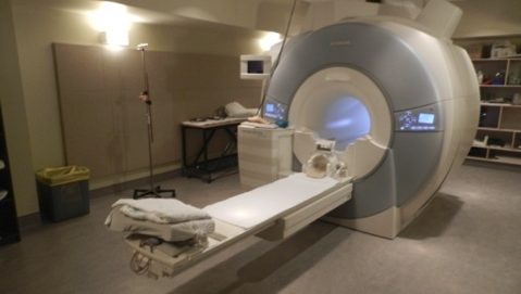
Douglas Brain Imaging Center (CIC)

Positron Emission Tomography (PET) is an imaging modality that produces metabolic processes’ representation of the body. Positron-emitting radioactive tracers are injected into the body, bind to biologically active molecules, such as glucose in the case of FDG-PET, and emit gamma rays that are detected by the scanner. Our lab uses one of the nineteen ECAT High-resolution Research Tomography (HRRT) scanners ever produced, dedicated for human PET scanner with LSO and GSO scintillators, allowing for photon detection with depth-of-interaction information. The ECAT HRRT has by far the highest resolution for a PET scanner. It contains 936 detectors, nearly four times the number found in ECAT EXACT HR+, its closest competitor.
Micro-PET can scan living animals in the same way PET is used in human studies. The Concorde R4 microPET is housed at the MNI, allowing for rodent (rat) imaging studies. This system consists of 32 rings of LSO detector crystal and 192 detectors, allowing for a resolution of 2mm. Imaging Alzheimer’s disease rat models using various metabolic tracers allows for better understanding of the disease. In our lab, we are using 18F-FDG PET to measure glucose metabolism (measuring neurodegeneration), 11C-PIB and 18F-NAV4694 to measure amyloid-beta (Aβ) plaques, and 18F-PBR06 tracer to measure neuro-inflammation. Douglas Brain Imaging Center (CIC).
The Douglas CIC is an imaging facility dedicated to conduct both preclinical and clinical imaging research. While human imaging studies typically use a 3-tesla MRI scanner, the Douglas CIC maintains a 7-tesla MRI Bruker System scanner for animal studies, allowing for high-resolution imaging. The CIC allows acts as a local platform for longitudinal studies in both humans and animals. In our lab, we conduct longitudinal analysis using these imaging modalities and other biomarkers in order to better understand the progression of AD. More information can be found at: http://www.douglas.qc.ca/page/bic.